 SCN 3D Atlas
About
The suprachiasmatic nucleus (SCN) is the master circadian pacemaker in mammals. SCN is a heterogeneous nucleus composed of a variety of cell types. These different cell types may have different spatial localizations in SCN. In the mouse, SCN was usually simply divided into two subdivisions: a ventral “core” region and a dorsal “shell” region. There was no physical boundary between core and shell but they can be distinguished by their neurochemical contents. Some studies also revealed the difference between anterior and posterior part of SCN. In order to study the spatial localizations of cell types in SCN, we introduce a 3D atlas of SCN.
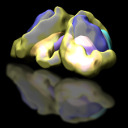
To image different cell types in SCN, cre line mice (Vip, Avp, Cck, Calb2, Calb1, Sst, Drd1, Gad2, vGlut2, Nms, Syt10, Rorb, Vipr2, Prokr2) were crossed to tdTomato reporter line (Ai9) such that specific types of cells were fluorescently labelled. Mice were sacrificed to harvest the thick brain slices containing the whole SCN. FRUIT method was used to achieve transparent brain slices. We also produced CLARITY immune fluorescence on SCN brain slices, in order to detect the spatial pattern of some neuropeptide that do not have good cre line, such as GRP. SCN region was imaged with confocal microscopes. With our newly developed computational method, every single neuron was detected in the confocal stack images and the information was saved into SWC files. SWC files from different experiments were integrated into a standard SCN atlas according to their DAPI signals in SCN. With the Neutube software, the neurons in each cell type were marked with individual color. We can also zoom in, zoom out, drag and drop to visualize them.